


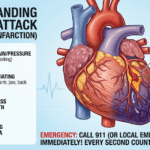





According to updated data from the American Heart Association (AHA) and the Centers for Disease Control and Prevention (CDC), cardiovascular disease remains the leading cause of mortality in the United States, accounting for nearly 1 in every 3 deaths. In fact, someone in the U.S. suffers a heart attack approximately every 40 seconds.
Despite these sobering metrics, modern cardiology has undergone a massive evolution. Discovering a heart issue early radically alters its trajectory. Today, the pathway to a heart disease diagnosis is highly precise, relying on a tiered combination of clinical history, predictive risk calculators, lab work, non-invasive monitoring, and advanced structural imaging.
If you or a loved one are scheduled for a cardiovascular health evaluation, understanding how specialists isolate specific heart conditions can demystify the process and alleviate clinical anxiety.
1. The Clinical Evaluation: The First Line of Defense
Every diagnostic journey begins long before you connect to a complex machine. A primary care physician or cardiologist always initiates the process with a thorough medical history and physical examination.
During this baseline evaluation, your care team maps out your individual risk profile. Under the latest medical guidelines, clinicians look closely at Cardiovascular-Kidney-Metabolic (CKM) syndrome risk factors, assessing how your heart interacts with your metabolic and renal health. They look closely at variables such as:
American Heart Association Journals
- Family History: Genetic predispositions to early-onset coronary artery disease (CAD), arrhythmias, or cardiomyopathy (enlarged heart muscle). American Heart Association
- Lifestyle Indicators: Tobacco use, sedentary habits, poor sleep quality, and dietary patterns. Healthline
- Comorbidities: Chronic conditions that damage blood vessels over time, specifically type 2 diabetes, chronic kidney disease, obesity, and sleep apnea. American Heart Association Journals
The clinical physical exam serves as an immediate, tactile assessment. By using a stethoscope to listen to your heart (auscultation), a physician can detect abnormal sounds like heart murmurs—which may indicate structural valve disorders—or an irregular rhythm. They will also measure your blood pressure and check for peripheral edema (swelling in the legs or ankles), a common physical indicator of congestive heart failure.
2. Risk Calculators and Blood Panels: Biomarkers in the Bloodstream
When heart tissue is under stress or damaged, it sheds specific proteins and enzymes into the blood. Blood tests provide an accessible, minimally invasive chemical snapshot of your cardiovascular profile.
The Shift to PREVENT Risk Scoring
In modern primary prevention, older risk scores have been replaced by a newer, more contemporary tool: the PREVENT (Predicting Risk of Cardiovascular Disease EVENTs) calculator. Unlike outdated models, the PREVENT equations accurately estimate both 10-year and 30-year risks of a heart attack or stroke for adults ages 30–79. It helps clinicians categorize risk into low (<3%), borderline (3% to <5%), intermediate (5% to <10%), and high (10% or higher) to personalize care earlier.
American Heart Association
Essential Blood Biomarkers
- Lipid Panel & Lipoprotein(a): Measures total cholesterol, HDL, and LDL. Medical updates strongly recommend measuring Lipoprotein(a) or Lp(a) at least once in adulthood. Because Lp(a) levels are genetically determined, a high level can double a person’s long-term risk of a heart attack or stroke, serving as a critical “hidden” risk factor. BaleDoneen+ 1
- High-Sensitivity CRP (hs-CRP): Evaluates systemic inflammation levels, which can signal the presence of unstable arterial plaques.
- Cardiac Troponin (T or I): Specific proteins released during myocardial injury. This is the gold standard for identifying acute heart attacks in progress within emergency departments. BaleDoneen
- B-Type Natriuretic Peptide (BNP): A hormone secreted by the heart ventricles when under high pressure. This marker is heavily utilized under global consensus definitions to identify early or asymptomatic stages of heart failure. BaleDoneen
3. Electrocardiogram (ECG/EKG): Tracking the Heart’s Electrical Circuit
If a physician suspects an issue based on your initial exam or labs, the next step is typically an Electrocardiogram (ECG or EKG).
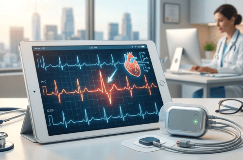
An ECG is a completely painless, non-invasive test that records the electrical signals traveling through your heart muscle. Small adhesive patches (electrodes) are placed on your chest, arms, and legs. Within minutes, the machine translates the electrical impulses into a series of wave patterns on a screen or strip of paper.
What an ECG Reveals:
- Heart Rate and Rhythm: Shows if the heart is beating too fast (tachycardia), too slow (bradycardia), or erratically (such as atrial fibrillation).
- Evidence of Prior Injury: Distorted wave patterns can reveal scars from a past, sometimes unnoticed, heart attack.
- Ischemia: Alterations in specific segments of the ECG graph can indicate that parts of the heart muscle are currently starved of oxygen.
Because a standard resting ECG only captures the heart’s behavior over a brief 10-second window, it may miss transient issues. If your signs of heart problems come and go unexpectedly, a specialist might prescribe a Holter Monitor or an Event Recorder. These are portable, wearable ECG devices that record your heart’s electrical patterns continuously for 24 hours up to 30 days while you go about your normal daily routine.
4. Echocardiogram: Real-Time Structural Ultrasound
While an ECG tests the heart’s internal electrical wiring, an Echocardiogram evaluates its mechanical pump design.
An echocardiogram utilizes high-frequency sound waves (ultrasound) to create live, moving images of your heart’s valves, chambers, and blood flow patterns. A technician glides a smooth wand (transducer) across your chest wall, projecting live data onto a viewing monitor.
Cardiologists rely heavily on a specific metric calculated during this test called the Ejection Fraction (EF).
Clinical Definition: Ejection Fraction is the percentage of blood pumped out of the heart’s main pumping chamber (the left ventricle) with each contraction.
Under the latest Universal Definition of Heart Failure, medical experts have shifted away from using rigid, static EF percentage cutoffs. Instead, heart failure is classified dynamically into categories: reduced, preserved, and improved EF. This reflects the clinical reality that a heart diagnosis is a moving trajectory; with early intervention and proper therapies, a weakened heart muscle can recover and improve over time.
American College of Cardiology
Beyond tracking pump strength, an echocardiogram can pinpoint thickening muscle walls (hypertrophy) or identify if any of the four cardiac valves are leaking (regurgitation) or failing to open fully (stenosis).
5. Stress Testing: Evaluating Function Under Exertion
Some cardiovascular vulnerabilities are completely invisible while you are resting comfortably on an exam table. They only manifest when the heart is forced to work harder, pumping faster to meet an increased demand for oxygen. This is the rationale behind the Cardiac Stress Test.
The standard protocol involves walking on a treadmill or riding a stationary bicycle while hooked up to an ECG monitor and blood pressure cuff. The speed and incline are increased at fixed intervals.
If physical mobility or musculoskeletal injuries prevent a patient from exercising safely, physicians can conduct a Pharmacological Stress Test. In this variant, intravenous medications (such as dobutamine or adenosine) are administered under strict supervision to safely mimic the physiological effects of strenuous exercise on the heart muscle.
Advanced Stress Testing Variants:
- Stress Echocardiogram: Ultrasound scans are performed immediately before and after exercise to check if specific sections of the heart wall fail to contract properly under stress—a sign of blocked coronary arteries.
- Nuclear Stress Test: A safe, tiny amount of a radioactive tracer is injected into the bloodstream. A specialized camera tracks the tracer to map out exactly how well blood flows through the heart muscle during exercise compared to rest. This helps spot areas with severe, chronic blockages.
6. Advanced Non-Invasive Cardiac Imaging
When standard tests point toward potential blockages or tissue abnormalities but leave lingering questions, specialized advanced imaging offers a deeper, highly detailed look.
+-----------------------------------+---------------------------------------------------------+
| Advanced Imaging Option           | Primary Diagnostic Benefit                              |
+-----------------------------------+---------------------------------------------------------+
| Coronary Artery Calcium (CAC) Scan| Measures calcified plaque; clarifies borderline/        |
|                                   | intermediate risk to guide statin therapy.      |
+-----------------------------------+---------------------------------------------------------+
| Coronary CT Angiography (CCTA)    | Uses contrast dye to construct 3D arterial models;      |
|                                   | catches dangerous soft plaques before they harden.       |
+-----------------------------------+---------------------------------------------------------+
| Cardiac MRI                       | Gold standard for tissue composition; distinguishes     |
|                                   | between active inflammation and permanent scars.        |
+-----------------------------------+---------------------------------------------------------+
A Coronary Artery Calcium (CAC) scan is highly recommended for men age 40 and older and women age 45 and older who fall into borderline or intermediate risk categories. If calcium buildup is present, it provides concrete evidence that supports setting stricter targets for lowering cholesterol to protect the arteries.
American Heart Association
7. Cardiac Catheterization: The Definitive Gold Standard
When non-invasive findings strongly suggest severe coronary artery blockages or complex structural defects, a specialist will schedule a Cardiac Catheterization, often paired with a Coronary Angiogram.
Unlike the previous methods, this is an invasive procedure, though it is routinely performed under light sedation. A long, thin, flexible tube called a catheter is carefully threaded through a blood vessel—typically starting at the wrist (radial artery) or the groin (femoral artery)—and guided safely up to the heart.
Once the catheter is positioned at the opening of the coronary arteries, a specialized contrast dye is released, and a continuous series of X-ray images (fluoroscopy) is taken.
This diagnostic procedure is uniquely powerful because it can instantly pivot into an immediate treatment window. If a cardiologist discovers a critically narrow artery during the diagnostic angiogram, they can perform an Angioplasty on the spot. They thread a tiny balloon through the catheter to open up the blockage, then deploy a small mesh tube called a stent to keep the artery safely propped open and restore normal blood flow.
Conclusion: Partnering with Your Healthcare Team
The path to a heart disease diagnosis is never a one-size-fits-all scenario. Your physician will deliberately select a sequence of diagnostic tools tailored to your unique symptoms, physical baseline, and personal risk profile.
If you are experiencing unexplained chest discomfort, shortness of breath during light activities, persistent dizziness, or heart palpitations, avoid waiting for symptoms to become severe. Reach out to a qualified healthcare professional promptly. Modern diagnostic methods can identify cardiovascular issues early with remarkable precision—empowering you and your medical team to protect your heart health well into the future.
American College of Cardiology
Disclaimer: This article is for informational purposes only and should not replace professional medical advice, diagnosis, or treatment. Always consult with your physician regarding any cardiovascular concerns.


![alt="8K hyper-realistic photograph inside a modern US electrophysiology clinic. A compassionate female cardiologist with PVC HEART branding on her lab coat points to a high-resolution monitor displaying an ECG tracing with an isolated wide QRS complex and compensatory pause. An attentive patient watches while holding a matching printed rhythm reference sheet. Soft ambient clinic lighting, medical precision, [MEDICALLY REVIEWED] trust seal visible."](https://pvcheart.com/wp-content/uploads/2026/07/watermarked_img_8726221352871066765-500x330.png)