






When experiencing unfamiliar chest flutters, brief pauses, or exertion-related fatigue, patients often struggle to understand what their heart is trying to communicate. Two common cardiac evaluations that frequently cross paths in clinical settings are Premature Ventricular Contractions (PVCs) and Heart Failure (HF).
Patients routinely ask: Do heart failure symptoms resemble PVCs? Could these benign-feeling extra beats actually be a sign of a failing heart muscle, or can frequent PVCs eventually cause heart failure?
Understanding the clinical intersection between these two distinct conditions requires evaluating electrical rhythm disturbances, structural heart muscle function, and how subtle mechanical deficits yield overlapping physical symptoms. Guidance provided by the American Heart Association Guidelines on Heart Failure Management emphasizes early diagnostic differentiation to protect long-term ventricular ejection fraction and prevent irreversible myocardial remodeling.
1. Electrical vs. Structural: The Fundamental Difference
To determine if symptoms overlap, it is vital to first differentiate the underlying pathology of each condition:
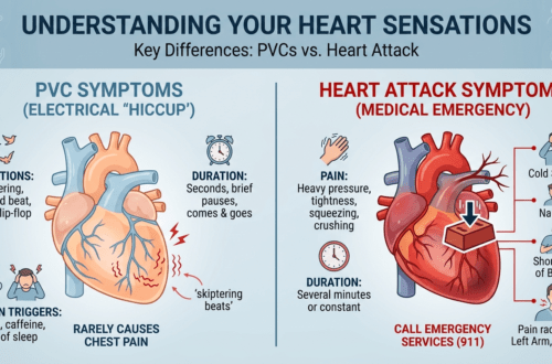
- Premature Ventricular Contractions (PVCs): An electrical rhythm anomaly. An irritable focus in the lower chambers (ventricles) fires ahead of schedule, causing an early heartbeat followed by a compensatory pause. In most cases, the underlying heart muscle remains structurally normal and strong.
- Heart Failure (HF): A structural/mechanical impairment where the heart muscle is either too weak (systolic failure / HFrEF) or too stiff (diastolic failure / HFpEF) to pump blood efficiently enough to meet the body’s metabolic demands.
       PREMATURE VENTRICULAR CONTRACTIONS                    HEART FAILURE
             (Electrical Issue)                          (Structural/Mechanical)
                     │                                              │
      Early Impulse in Ventricle                     Weak or Stiff Heart Muscle
                     │                                              │
        Intermittent Beat Pause                      Reduced Systemic Blood Flow
                     │                                              │
    "Flutter, Flip-Flop, or Hard Thump"          "Fluid Retention & Exertional Fatigue"
While one is primarily an electrical timing error and the other is a mechanical pumping deficiency, their clinical presentations frequently intersect in patient-reported symptoms.
2. Do Heart Failure Symptoms Resemble PVCs? Where They Overlap
The answer is yes—under specific circumstances, heart failure symptoms can closely resemble PVCs, and vice versa. Because an ectopic beat represents an inefficient cardiac contraction (pumping significantly less blood than a normal beat), frequent PVCs can temporarily reduce cardiac output, triggering physical symptoms almost identical to early-stage heart failure.
┌─────────────────────────────────────────────────────────────────────────────┐
│                    OVERLAPPING SYMPTOM MANIFESTATIONS                       │
├──────────────────────────┬──────────────────────────────────────────────────┤
│ Symptom                  │ Why It Occurs in Both Conditions                 │
├──────────────────────────┼──────────────────────────────────────────────────┤
│ **Shortness of Breath    │ • PVCs: Sudden loss of effective stroke volume   │
│ (Dyspnea)**              │   during high-frequency ectopic runs.            │
│                          │ • HF: Fluid congestion backing up into lungs.    │
├──────────────────────────┼──────────────────────────────────────────────────┤
│ **Fatigue & Lethargy**   │ • PVCs: Chronic rhythm interruption reduces peak │
│                          │   cardiac output.                                │
│                          │ • HF: Inadequate oxygenated blood to muscles.    │
├──────────────────────────┼──────────────────────────────────────────────────┤
│ **Palpitations & Chest   │ • PVCs: Sudden forceful beat following a pause.  │
│ Awareness**              │ • HF: Compensatory tachycardia or secondary      │
│                          │   arrhythmias triggered by chamber stretch.      │
└──────────────────────────┴──────────────────────────────────────────────────┘
Detailed physiological models detailing reduced stroke volume and altered hemodynamics during arrhythmia events are maintained by the National Library of Medicine (NCBI) Arrhythmia Research Database.
3. Symptom Comparison Matrix: PVCs vs. Heart Failure
While symptoms can overlap, key clinical characteristics separate isolated, benign ectopic beats from progressive heart failure.
| Clinical Presentation | Isolated / Benign PVCs | Structural Heart Failure |
|---|---|---|
| Primary Sensation | “Skipped beat,” “flip-flop,” or sudden hard “thump” in the chest. | Constant chest heaviness, breathlessness, and profound body fatigue. |
| Timing of Symptoms | Most noticeable at rest, during relaxation, or when lying down in bed. | Worsens progressively during physical exertion or exercise. |
| Fluid Retention | None. No swelling in limbs or rapid weight gain. | Presence of peripheral edema (swollen ankles, feet, legs) and abdominal bloating. |
| Positional Breathing | Breathing remains normal when lying flat on your back. | Orthopnea: Shortness of breath when lying flat, requiring elevated pillows. |
| Onset Profile | Instantaneous, episodic, coming and going in seconds or minutes. | Chronic, steadily worsening over weeks or months. |
4. The Two-Way Street: PVC-Induced Cardiomyopathy vs. HF-Triggered PVCs
The relationship between PVCs and heart failure is not merely a diagnostic coincidence; it is a dynamic clinical spectrum where one can directly cause or worsen the other.
┌─────────────────────────────────────────────────────────────────────────────┐
│                       THE PVC-HEART FAILURE CYCLE                           │
├─────────────────────────────────────────────────────────────────────────────┤
│                                                                             │
│  Option A:  Frequent PVCs (>10-15%) ──► Dyssynchrony ──► PVC Cardiomyopathy │
│                                                                 │           │
│                                                                 ▼           │
│  Option B:  Heart Failure ──► Chamber Stretch & Scar ──► Secondary PVCs     │
│                                                                             │
└─────────────────────────────────────────────────────────────────────────────┘
1. PVC-Induced Cardiomyopathy (Ectopy Causing Heart Failure)
When a patient experiences a high PVC burden—meaning extra beats account for 10% to 15% or more of their total daily heartbeats—the ventricles contract asynchronously for months or years.
This uncoordinated mechanical squeezing can cause the left ventricle to dilate and weaken, leading to a reversible form of heart failure known as PVC-induced cardiomyopathy. In these cases, suppressing or eliminating the extra beats via pharmacological therapy or radiofrequency catheter ablation often completely restores heart function. Clinical studies archived in the NCBI Medical Literature Repository demonstrate significant recovery in ejection fraction following successful ablation of high-burden ectopic foci.
2. Heart Failure Triggering Ectopic Beats
Conversely, pre-existing heart failure stretches the muscle tissue in the lower chambers. This structural remodeling, combined with elevated adrenaline levels and altered cellular electrolytes, creates localized “irritable spots” in the ventricular walls, making ectopic beats far more frequent.
According to epidemiological data published by the Centers for Disease Control and Prevention (CDC) Heart Disease Division, patients with underlying structural heart disease or heart failure frequently exhibit complex ectopic activity requiring routine ambulatory monitoring and risk stratification.
5. Diagnostic Workup: Distinguishing the Root Cause
To accurately differentiate between isolated ectopic beats, underlying heart failure, or a combination of both, cardiologists use a standardized diagnostic sequence:
┌──────────────────────────────────────────────────────────────────────────┐
│                   DIAGNOSTIC WORKUP FOR PVC/HF EVALUATION                │
├───────────────────┬───────────────────┬──────────────────────────────────┤
│ Diagnostic Test   │ Primary Purpose   │ Target Findings                  │
├───────────────────┼───────────────────┼──────────────────────────────────┤
│ **12-Lead ECG**   │ Captures exact    │ Identifies QRS morphology, baseline│
│                   │ ectopic waveforms │ intervals, and acute ischemia.   │
├───────────────────┼───────────────────┼──────────────────────────────────┤
│ **Holter Monitor**│ Measures 24-48 hr │ Calculates exact % ectopic burden│
│                   │ rhythm frequency  │ and unifocal vs. multifocal runs.│
├───────────────────┼───────────────────┼──────────────────────────────────┤
│ **Transthoracic   │ Evaluates heart   │ Measures Left Ventricular        │
│ Echocardiogram**  │ structure/function│ Ejection Fraction (LVEF) & valves.│
├───────────────────┼───────────────────┼──────────────────────────────────┤
│ **NT-proBNP Blood │ Assesses cardiac  │ Elevated peptide levels indicate  │
│ Test**            │ wall stress       │ active heart failure strain.     │
└───────────────────┴───────────────────┴──────────────────────────────────┘
Calculating Ejection Fraction (LVEF)
The most definitive boundary between isolated PVCs and heart failure is the Left Ventricular Ejection Fraction (LVEF), measured during an echocardiogram:
LVEF (%)=(End-Diastolic VolumeEnd-Diastolic Volume−End-Systolic Volume)×100
- Normal LVEF (50%−70%): Heart muscle is pumping effectively. PVCs are typically benign.
- Reduced LVEF (<40%): Indicates heart failure with reduced ejection fraction (HFrEF), requiring formal medical therapy as recommended by the American College of Cardiology (ACC) Diagnostic Standards.
6. Red Flag Symptoms: When to Seek Immediate Medical Care
While isolated premature beats are generally harmless in healthy hearts, specific symptoms warrant urgent medical evaluation to rule out acute heart failure decompensation or malignant arrhythmias:
- Shortness of breath that wakes you up at night (Paroxysmal Nocturnal Dyspnea).
- Rapid, unexplained weight gain (e.g., 2−3 lbs in 24 hours or 5 lbs in a week due to fluid retention).
- Swelling in both feet, ankles, legs, or abdomen (Pitting Edema).
- Episodes of near-fainting, lightheadedness, or full loss of consciousness (Syncope).
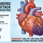
- Chest pain, pressure, or tightness accompanying rhythm irregularities.
Key Diagnostic Terms Reference
When reviewing medical reports or discussing results with your care team, refer to this clinical terminology table:
| Medical Term | Plain Language Meaning | Clinical Relevance |
|---|---|---|
| Ejection Fraction (EF) | Percentage of blood pumped out of the main chamber with each beat. | Primary metric used to diagnose heart failure. |
| Ectopic Burden | Percentage of daily heartbeats that are premature beats. | Burdens above 10−15% can cause reversible muscle weakening. |
| NT-proBNP / BNP | Hormone released by heart muscle when under excess pressure/stretch. | Blood marker used to detect active heart failure strain. |
| Cardiomyopathy | Disease of the heart muscle that impairs pumping ability. | Can be the cause or the result of frequent ectopic beats. |
| Orthopnea | Difficulty breathing while lying flat. | Classic symptom of heart failure fluid congestion. |
Medical Disclaimer
Disclaimer: The content provided on PVC Heart, including text, graphics, images, and other material, is for informational and educational purposes only. It is not intended to be a substitute for professional medical advice, diagnosis, or treatment. Always seek the advice of your physician, cardiologist, or electrophysiologist with any questions you may have regarding heart rhythm symptoms, heart failure, or diagnostic testing. Never disregard professional medical advice or delay in seeking it because of something you have read on this website. If you experience severe shortness of breath, chest pain, or fainting, call 911 or visit the nearest emergency room immediately.