








Premature Ventricular Contractions (PVCs) are extra heartbeats. They start in your heart’s lower chambers (ventricles). These extra beats disrupt your regular heart rhythm. You might feel a flutter, a flip-flop, or a skipped beat in your chest.
Most PVCs are harmless. However, frequent PVCs need proper medical evaluation. Getting an accurate premature ventricular contractions diagnosis helps doctors find the cause and protect your long-term heart health.
This guide explains how cardiologists diagnose PVCs in the United States, what tests you will take, and what your results mean.
Initial Medical Evaluation
Your diagnostic journey starts in a clinic. A doctor or cardiologist will evaluate your health history and physical symptoms.
Physical Examination
During your physical exam, the doctor will:
- Listen to your heart with a stethoscope.
- Check your pulse rate and rhythm.
- Measure your blood pressure.
- Look for signs of fluid retention or thyroid issues.
Sometimes, an extra heartbeat occurs right during your exam. Your doctor can hear or feel this irregular pattern directly.
Medical History and Symptoms Review
Your provider will ask detailed questions about your daily life. Be ready to share:
- How often you feel heart palpitations.
- When symptoms usually occur (at rest, during stress, or after exercise).
- Your daily caffeine, alcohol, and nicotine intake.
- Current prescription medications and over-the-counter supplements.
- Your personal or family history of heart disease.
Primary Diagnostic Tests for PVCs
Several non-invasive tests help identify and quantify premature beats.
1. Electrocardiogram (ECG or EKG)
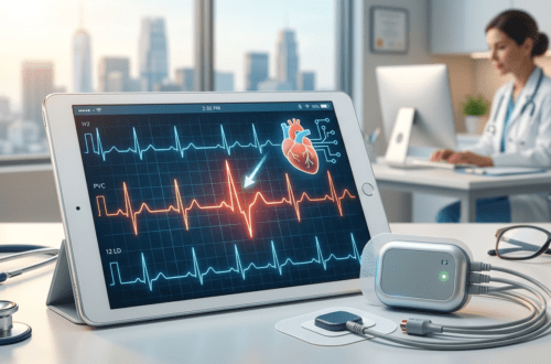
An ECG is the main tool used to diagnose PVCs. It records the electrical activity of your heart using small sticky patches (electrodes) placed on your chest, arms, and legs.
- How it works: The machine traces your heart’s electrical signals onto paper or a screen.
- What it shows: PVCs produce a distinct, wide electrical pattern on the tracing.
- Limitations: A standard ECG only lasts about 10 seconds. If you do not have a PVC during those 10 seconds, the test will look completely normal.
Because standard ECGs are brief, doctors often use continuous monitoring devices.
Ambulatory Heart Monitors
If your standard ECG is clear but you still have symptoms, your cardiologist will order portable monitoring.
Holter Monitor
A Holter monitor is a small, wearable ECG device. You wear it continuously for 24 to 48 hours while going about your normal daily activities.
- Purpose: It records every single heartbeat over 1 to 2 days.
- Benefit: It determines your PVC burden. PVC burden is the percentage of your total daily heartbeats that are PVCs.
- Logbook: You keep a diary of your activities and symptoms so your doctor can match your feeling to the recording.
Event Monitor
If your symptoms happen infrequently—such as once or twice a week—a Holter monitor might miss them. An event monitor is worn for up to 30 days.
- Symptom-Triggered: You press a button to record electrical signals only when you feel a flutter.
- Auto-Triggered: Some models automatically start recording whenever an abnormal rhythm occurs.
Patch Monitors
Modern patch monitors are lightweight, wire-free adhesive pads worn on the chest. They record data for up to two weeks, providing a comfortable option for extended tracking.
Testing for Underlying Heart Conditions
Diagnosing PVCs is only the first step. Doctors must also check if an underlying structural heart problem is triggering these extra beats.
| Test Type | Primary Purpose | What It Measures |
| Echocardiogram | Ultrasound imaging | Heart structure, valve function, and ejection fraction |
| Stress Test | Exercise monitoring | Heart rhythm response to physical strain |
| Cardiac MRI | Detailed tissue imaging | Microscopic heart scarring or subtle muscle damage |
| Blood Tests | Chemical analysis | Electrolytes, thyroid hormones, and kidney function |
Echocardiogram (Echo)
An echocardiogram uses sound waves to create live pictures of your heart. It evaluates:
- The size and shape of your heart chambers.
- How well your heart pumps blood (ejection fraction).
- Valve movement and structural heart muscle issues.
A normal echocardiogram usually confirms that your PVCs are benign (harmless).
Exercise Stress Test
A stress test monitors your ECG while you walk on a treadmill or ride a stationary bike.
- If PVCs disappear during exercise: This often indicates a benign condition.
- If PVCs increase during exercise: This suggests that adrenaline or stress triggers your arrhythmia, which may require medical management.
Cardiac MRI or CT Scan
If your doctor suspects subtle heart muscle damage, inflammation (myocarditis), or scarring, they may order a Cardiac MRI. This provides high-resolution images of heart tissue.
Laboratory Tests and Secondary Causes
Sometimes PVCs originate outside the heart. Blood work helps uncover systemic triggers.
Key blood tests include:
- Electrolyte Panel: Checks levels of potassium, magnesium, and calcium. Low levels can irritate heart muscle cells.
- Thyroid Function Tests: Measures thyroid hormone activity. An overactive thyroid (hyperthyroidism) often triggers extra heartbeats.
- Complete Blood Count (CBC): Checks for anemia, which forces the heart to work harder.
Understanding Your PVC Burden
Once diagnostic testing is complete, your cardiologist calculates your total PVC burden.
- Low Burden (Less than 1%): Less than 1,000 PVCs per day. Typically benign and rarely requires medication unless symptoms are severe.
- Moderate Burden (1% to 10%): 1,000 to 10,000 PVCs per day. Requires periodic monitoring to ensure heart strength remains normal.
- High Burden (Greater than 10% to 15%): More than 10,000 to 15,000 PVCs per day. Over time, a high burden can weaken the heart muscle (PVC-induced cardiomyopathy). Treatment is strongly recommended.
When to Seek Immediate Medical Evaluation
Most PVCs are harmless, but certain symptoms require urgent attention. Contact a doctor immediately or visit an emergency room if your heart flutters are accompanied by:
- Chest pain, pressure, or tightness.
- Severe shortness of breath.
- Dizziness, lightheadedness, or fainting (syncope).
- A history of structural heart disease or heart attack.
Summary
A proper premature ventricular contractions diagnosis combines a physical exam, rhythm tracking (ECG and Holter monitoring), and structural imaging (Echocardiogram). Identifying your PVC burden and ruling out underlying heart conditions gives you and your doctor a clear path forward for effective management and peace of mind.Rread more
![alt="8K hyper-realistic photograph inside a modern US medical clinic. A reassuring female cardiologist (PVC HEART branding, Dr. Sarah Jenkins) is seated at a desk, softly pointing to a wall monitor that explicitly displays the title 'BLOOD WORK FOR HEART PALPITATIONS: KEY LAB TESTS' and structured data for electrolytes, thyroid, CBC, and glucose. An attentive American patient listens, holding the matching printed checklist. Natural golden hour lighting, sharp detail, [MEDICALLY REVIEWED] seal visible."](https://pvcheart.com/wp-content/uploads/2026/07/Gemini_Generated_Image_catzaucatzaucatz-75x75.png)

![alt="8K hyper-realistic photograph inside a modern US electrophysiology clinic. A compassionate female cardiologist with PVC HEART branding on her lab coat points to a high-resolution monitor displaying an ECG tracing with an isolated wide QRS complex and compensatory pause. An attentive patient watches while holding a matching printed rhythm reference sheet. Soft ambient clinic lighting, medical precision, [MEDICALLY REVIEWED] trust seal visible."](https://pvcheart.com/wp-content/uploads/2026/07/watermarked_img_8726221352871066765-500x330.png)