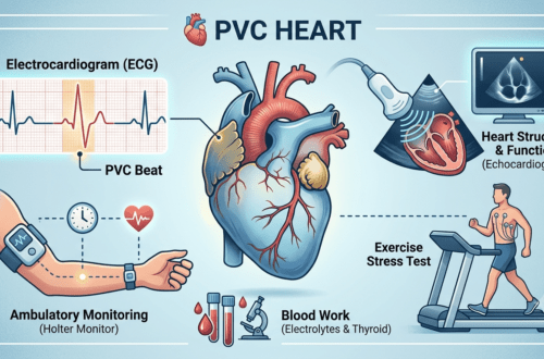






Experiencing a sudden heart flutter, a heavy thump in your chest, or a sensation that your heart skipped a beat can be alarming. When patients report these symptoms, the diagnostic journey usually starts with an electrocardiogram (ECG/EKG) or wearable continuous cardiac monitor. For more on these first-line tools, see the American Heart Association (AHA) Guide to Heart Palpitations.
However, many cardiac palpitations are not caused by a primary structural problem within the heart itself. Instead, they are often secondary responses triggered by systemic metabolic shifts, endocrine imbalances, or nutrient deficiencies.
Targeted blood work acts as a foundational screening tool to identify underlying systemic triggers. Here is an evidence-based breakdown of the essential blood tests your healthcare provider should consider ordering to pinpoint non-cardiac causes of heart rhythm disturbances.
1. Electrolyte Panel: The Fuel for Cardiac Conduction
Your heart cells rely on electrically charged minerals—electrolytes—to generate and transmit the electrical signals that govern every single heartbeat. As detailed in the National Library of Medicine (NCBI) Electrolyte Panel Overview, subtle deviations in serum concentrations can destabilize myocardial electrical activity.
[ Normal Electrolyte Levels ] ──► Steady Electrical Impulses ──► Regular Heartbeat
[ Low Potassium / Magnesium ] ──► Irritated Cardiac Tissue  ──► Palpitations / Ectopy
Serum Potassium ($K^+$)
Potassium plays a direct role in resting cardiac membrane potential and repolarization.
- Hypokalemia (Low Potassium): Low potassium increases myocardial excitability, frequently leading to premature ectopic beats or fast atrial/ventricular rhythms. Common causes include diuretic medications, excessive sweating, or gastrointestinal issues.
- Hyperkalemia (High Potassium): High potassium alters electrical conduction velocity, which can cause dangerous cardiac arrhythmias or conduction blocks.
Serum Magnesium ($Mg^{2+}$)
Magnesium is a critical cofactor for the sodium-potassium pump ($Na^+/K^+$-ATPase) that maintains cellular electrical balance.
- Because serum magnesium tests account for less than 1% of total body magnesium stores, even a “low-normal” result can reflect a tissue deficiency that irritates cardiac muscle cells.
- Low magnesium frequently co-occurs with low potassium, making it difficult to correct potassium levels without restoring magnesium first.
Serum Calcium ($Ca^{2+}$)
Calcium governs the physical contraction mechanism of the heart muscle. Both hypocalcemia (low calcium) and hypercalcemia (high calcium) alter the heart’s QT interval on an ECG, predisposing patients to irregular rhythms or palpitations.
2. Complete Blood Count (CBC): Oxygen Delivery & Systemic Stress
A Complete Blood Count (CBC) provides a detailed snapshot of your circulating blood cells and is a standard part of diagnostic safety protocols established by the CDC Division of Laboratory Systems.
Hemoglobin and Hematocrit (Anemia Screening)
Red blood cells transport oxygen from your lungs to your body’s tissues. When red blood cell counts or hemoglobin levels drop (anemia), tissue oxygen delivery decreases. To compensate and prevent tissue hypoxia:
- The sympathetic nervous system releases epinephrine (adrenaline).
- The heart compensates by pumping faster (tachycardia) and striking harder.
- This compensatory mechanism is experienced by the patient as a pounding, racing, or fluttering sensation in the chest.
Serum Ferritin and Iron Panel
Beyond a basic CBC, testing ferritin (the body’s iron storage protein) is crucial. Patients can suffer from functional iron deficiency even before overt anemia develops on a CBC, which can still trigger hyperdynamic circulation and cardiac symptoms.
3. Comprehensive Thyroid Panel: The Body’s Metabolic Thermostat
The thyroid gland secretes hormones that directly regulate cardiac output, heart rate, and vascular resistance. Thyroid dysfunction is one of the most frequent endocrine causes of heart palpitations.
┌─────────────────────────────────────────────────────────────────────────┐
│                      THYROID FUNCTION & THE HEART                        │
├───────────────────────────────────┬─────────────────────────────────────┤
│ Hyperthyroidism (Overactive)     │ Hypothyroidism (Underactive)        │
├───────────────────────────────────┼─────────────────────────────────────┤
│ • Elevated Free T3 / Free T4      │ • Low Free T3 / Free T4             │
│ • Suppressed TSH levels           │ • Elevated TSH levels               │
│ • Increases SA node sensitivity   │ • Slows heart rate (bradycardia)    │
│ • Causes racing heart & ectopy    │ • Causes hard, heavy thumping beats │
└───────────────────────────────────┴─────────────────────────────────────┘
When evaluating heart rhythm issues, a complete panel should include:
- Thyroid-Stimulating Hormone (TSH): The primary screening marker released by the pituitary gland.
- Free Thyroxine ($FT_4$) and Free Triiodothyronine ($FT_3$): The active unbound circulating thyroid hormones. Checking active hormone levels ensures subclinical thyroid issues are not missed if TSH alone appears borderline.
4. Metabolic Health: Fasting Blood Glucose and HbA1c
Fluctuations in blood sugar trigger neuroendocrine responses that directly impact heart rhythm.
- Hypoglycemia (Low Blood Sugar): When blood glucose drops sharply, the adrenal glands release counter-regulatory hormones—specifically adrenaline and cortisol. This emergency surge causes a rapid heart rate, tremors, sweating, and strong palpitations.
- Hyperglycemia and Insulin Resistance: Chronic high blood sugar damages the autonomic nerves that control involuntary heart rate regulation (autonomic neuropathy), making the heart more susceptible to baseline rhythm disturbances.
Recommended screenings include a Fasting Plasma Glucose test and a Hemoglobin A1c (HbA1c) test to evaluate average glucose control over the preceding 3 months.
5. Inflammatory Markers & Cardiac Biomarkers
In select clinical cases—particularly if palpitations start suddenly following an infection or are accompanied by shortness of breath or fatigue—specialized markers are warranted:
- High-Sensitivity C-Reactive Protein (hs-CRP): Measures systemic vascular inflammation. Low-grade inflammation can irritate the pericardium (heart lining) or myocardium (heart muscle).
- High-Sensitivity Cardiac Troponin: Released into the bloodstream only when heart muscle cells experience cellular stress or injury. Troponin tests help rule out active myocardial inflammation (myocarditis) or ischemic events.
- B-Type Natriuretic Peptide (BNP / NT-proBNP): A hormone secreted by the heart ventricles when subjected to increased pressure or stretch. Elevated levels suggest the heart muscle is working under strain, aiding in the early detection of heart failure or structural overload.
Key Lab Panel Checklist for Your Next Appointment
When preparing for your medical consultation, print or save this summary checklist to review with your provider:
| Lab Category | Specific Tests to Request | What It Helps Rule Out |
| Electrolyte Panel | Potassium, Magnesium, Calcium, Sodium | Electrical conduction instability & membrane irritability |
| Hematology | Complete Blood Count (CBC), Serum Ferritin | Anemia, impaired oxygen delivery, & iron deficiency |
| Endocrine | TSH, Free $T_3$, Free $T_4$ | Hyperthyroidism & metabolic rate acceleration |
| Glycemic Control | Fasting Glucose, HbA1c | Adrenaline surges from low sugar or diabetic autonomic changes |
| Cardiac Biomarkers | High-Sensitivity Troponin, NT-proBNP | Myocardial injury, active inflammation, or heart strain |
Medical Disclaimer
Disclaimer: The content provided on PVC Heart, including text, graphics, images, and other material, is for informational and educational purposes only. It is not intended to be a substitute for professional medical advice, diagnosis, or treatment. Always seek the advice of your physician, cardiologist, or other qualified healthcare provider with any questions you may have regarding a medical condition or lab results. Never disregard professional medical advice or delay in seeking it because of something you have read on this website. If you think you may have a medical emergency, call 911 or your local emergency services immediately.
![alt="8K hyper-realistic photograph inside a modern US electrophysiology clinic. A compassionate female cardiologist with PVC HEART branding on her lab coat points to a high-resolution monitor displaying an ECG tracing with an isolated wide QRS complex and compensatory pause. An attentive patient watches while holding a matching printed rhythm reference sheet. Soft ambient clinic lighting, medical precision, [MEDICALLY REVIEWED] trust seal visible."](https://pvcheart.com/wp-content/uploads/2026/07/watermarked_img_8726221352871066765-75x75.png)

![alt="8K hyper-realistic photograph inside a modern US electrophysiology clinic. A compassionate female cardiologist with PVC HEART branding on her lab coat points to a high-resolution monitor displaying an ECG tracing with an isolated wide QRS complex and compensatory pause. An attentive patient watches while holding a matching printed rhythm reference sheet. Soft ambient clinic lighting, medical precision, [MEDICALLY REVIEWED] trust seal visible."](https://pvcheart.com/wp-content/uploads/2026/07/watermarked_img_8726221352871066765-500x330.png)